Evolutionists fancifully forges so many theories and wishful thinking as to how a organism 'evolved' from a simpler state, development to a fully formed one.. But they don't explain the details, surely because they make the seeming probability of evolution occurring even smaller (supposing that it actually had any probability to occur at all!)
Darwin himself found amazing and quite improbable for a eye, to have evolved through minute developments, improvement, during millions of years.. Let alone if he knew about that:
Biochemical sketch of
the eye's operation:

When light first strikes the retina a photon interacts with a molecule
called 11-cis-retinal, which rearranges within picoseconds to trans-retinal.
(A picosecond is about the time it takes light to
travel the breadth of a single human hair.) The change in the shape of
the retinal molecule forces a change in the shape of the protein, rhodopsin,
to which the retinal is tightly bound. The protein's metamorphosis alters
its behavior. Now called metarhodopsin II, the protein sticks to another
protein, called transducin. Before bumping into metarhodopsin II,
transducin had tightly bound a small molecule called GDP. But when
transducin interacts with metarhodopsin II, the GDP falls off, and a
molecule called GTP binds to
transducin. (GTP is closely related to, but critically different from, GDP.)
GTP-transducin-metarhodopsin II now binds to a protein called
phosphodiesterase, located in the inner membrane of the cell. When
attached to metarhodopsin II and its entourage, the phosphodiesterase
acquires the chemical ability to «cut» a molecule called cGMP (a chemical
relative of both GDP and GTP). Initially there are a lot of cGMP molecules
in the cell, but the phosphodiesterase lowers its concentration, just as a
pulled plug lowers the water level in a bathtub.
Another membrane protein that binds cGMP is called an ion channel.
It acts as a gateway that regulates the number of sodium ions in the cell.
Normally the ion channel allows sodium ions to flow into the cell, while
a separate protein actively pumps them out again. The dual action of the
ion channel and pump keeps the level of sodium ions in the cell within a
narrow range. When the amount of cGMP is reduced because of cleavage
by the phosphodiesterase, the ion channel closes, causing the cellular
concentration of positively charged sodium ions to be reduced. This causes
an imbalance of charge across the cell membrane that, finally, causes a
current to be transmitted down the optic nerve to the brain. The result,
when interpreted by the brain, is vision.
If the reactions mentioned above were the only ones that operated in
the cell, the supply of 11-cis-retinal, cGMP and sodium ions would quickly
be depleted. Something has to turn off the proteins that were turned on
and restore the cell to its original state. Several mechanisms do this. First,
in the dark the ion channel (in addition to sodium ions) also lets calcium
ions into the cell. The calcium is pumped back out by a different protein so
that a constant calcium concentration is maintained. When cGMP levels
fall, shutting down the ion channel, calcium ion concentration decreases,
too. The phosphodiesterase enzyme, which destroys cGMP slows down at
lower calcium concentration. Second, a protein called guanylate cyclase
begins to resynthesize cGMP when calcium levels start to fall. Third,
while all of this is going on, metarhodopsin II is chemically modified by
an enzyme called rhodopsin kinase. The modified rhodopsin then binds to
a protein known as arrestin, which prevents the rhodopsin from activating
more transducin. So the cell contains mechanisms to limit the amplified
signal started by a single photon.
Trans-retinal eventually falls off of rhodopsin and must be reconverted
to 11-cis-retinal and again bound by rhodopsin to get back to the starting
point for another visual cycle. To accomplish this, trans-retinal is first
chemically modified by an enzyme to trans-retinol—a form containing
two more hydrogen atoms. A second enzyme then converts the molecule
to 11-cis-retinol. Finally, a third enzyme removes the previously added
hydrogen atoms to form 11-cis-rennal, a cycle is complete. .
And that...
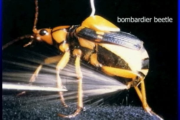
BEETLE BOMBS
The bombardier beetle is an insect of unassuming appearance,
measuring about one half-inch in length. When it is threatened by another
bug, however, the beetle has a special method of defending itself, squirting
a boiling-hot solution at the enemy out of an aperture in its hind section.16
The heated liquid scalds its target, which then usually makes other plans
for dinner. How is this trick done?
It turns out that the bombardier beetle is using chemistry. Prior to battle,
specialized structures called secretory lobes make a very concentrated
mixture of two chemicals, hydrogen peroxide and hydroquinone.
The hydrogen peroxide is the same material as one can buy in a
drugstore; hydroquinone is used in photographic development. The
mixture is sent into a storage chamber called the collecting vesicle.
The collecting vesicle is connected to, but ordinarily sealed off from, a
second compartment called (evocatively) the explosion chamber. The
two compartments are kept separate from one another by a duct with a
sphincter muscle, much like the sphincter muscles upon which humans
depend for continence. Attached to the explosion chamber are a number
of small knobs called ectodermal glands; these secrete enzyme catalysts
into the explosion chamber. When the beetle feels threatened it squeezes
muscles surrounding the storage chamber while simultaneously relaxing
the sphincter muscle. This forces the solution of hydrogen peroxide and
hydroquinone to enter the explosion chamber, where it mixes with the
enzyme catalysts.

Now, chemically, things get very interesting. The hydrogen peroxide
rapidly decomposes into ordinary water and oxygen, just as a store-bought
bottle of hydrogen peroxide will decompose over time if left open. The
oxygen reacts with the hydroquinone to yield more water, plus a highly
irritating chemical called quinone. These reactions release a large quantity
of heat. The temperature of the solution rises to
the boiling point; in fact, a portion vaporizes into steam. The steam
and oxygen gas exert a great deal of pressure on the walk of the explosion
chamber. With the sphincter muscle now closed, a channel leading outward
from the beetle's body provides the only exit for the boiling mixture.
Muscles surrounding the channel allow the steam jet to be directed at the
source of danger. The end result is that the beetle's enemy is scalded by a
steaming solution of the toxic chemical quinone.
You may wonder why the mixture of hydrogen peroxide and quinone
did not react explosively when they were in the collecting vesicle. The
reason is that many chemical reactions occur quite slowly if there is no
easy way for the molecules to get together on the atomic level—otherwise,
this book would burst into flame as it reacted with oxygen in the air. As
an analogy, consider a locked door. There is no easy way for people (say,
teenage boys and girls) on opposite sides of the door to get together, even
if they would be happy to do so. If someone has the key, however, then the
door can be opened and proper introductions can be made. The enzyme
catalysts play the role of the key, allowing the hydrogen peroxide and hydroquinone to get together
on the atomic level so that a reaction can take place.
The cascade
The body commonly stores enzymes (proteins that catalyze a chemical
reaction, like the cleavage of fibrinogen) in an inactive form for later use.
The inactive forms are called proenzymes. When a signal is received that
a certain enzyme is needed, the corresponding proenzyme is activated to
give the mature enzyme. As with the conversion of fibrinogen to fibrin,
proenzymes are often activated by cutting off a piece of the proenzyme
that is blocking a critical area. The strategy is commonly used with
digestive enzymes. Large quantities can be stored as inactive proenzymes,
then quickly activated when the next good meal comes along.
Thrombin initially exists as the inactive form, prothrombin. Because it
is inactive, prothrombin can't cleave fibrinogen, and the animal is saved
from death by massive, inappropriate clotting. Still, the dilemma of control
remains. If the cartoon saw were inactivated, the telephone pole would not
fall at the wrong time. If nothing switches on the saw, however, then it
would never cut the rope; the pole wouldn't fall even at the right time. If
fibrinogen and prothrombin were the only proteins in the blood-clotting
pathway, again our animal would be in bad shape. When the animal
was cut, prothrombin would just float helplessly by the fibrinogen as the
animal bled to death. Because prothrombin cannot cleave fibrinogen to
fibrin, something is needed to activate prothrombin.
Perhaps the reader can see why the blood-clotting system is called a
cascade—a system where one component activates another component,
which activates a third component, and so on.
A protein called Stuart factor cleaves prothrombin, turning it into active
thrombin that can then cleave fibrinogen to fibrin to form the blood clot.
Unfortunately, as you may have guessed, if Stuart factor, prothrombin,
and fibrinogen were the only blood-clotting proteins, then Stuart factor
would rapidly trigger the cascade, congealing all the blood of the organism. So Stuart factor also exists in an inactive
form that must first be activated.
At this point there's a little twist to our developing chicken-and-egg
scenario. Even activated Stuart factor can't turn on prothrombin. Stuart
factor and prothrombin can be mixed in a test tube for longer than it would
take a large animal to bleed to death without any noticeable production
of thrombin. It turns out that another protein, called accelerin, is needed
to increase the activity of Stuart factor. The dynamic duo—accelerin
and activated Stuart factor— cleave prothrombin fast enough to do the
bleeding animal some good. So in this step we need two separate proteins
to activate one proenzyme.
Yes, accelerin also initially exists in an inactive form, called proaccelerin
(sigh). And what activates it? Thrombin! But thrombin, as we have seen,
is further down the regulatory cascade than proaccelerin. So thrombin
regulating the production of accelerin is like having the granddaughter
regulate production of the grandmother. Nonetheless, due to a very low
rate of cleavage of prothrombin by Stuart factor, it seems there is always
a trace of thrombin in the bloodstream. Blood clotting is therefore auto-
catalytic, because proteins in the cascade accelerate the production of
more of the same proteins.
We need to back up a little at this point because, as it turns out,
prothrombin as it is initially made by the cell can't be transformed into
thrombin, even in the presence of activated Stuart factor and accelerin.
Prothrombin must first be modified by having
ten specific amino acid residues, called glutamate (Glu) residues, changed
to «.-carboxyglutamate (Gla) residues. The modification can be compared
to placing a lower jaw onto the upper jaw of a skull. The completed
structure can bite and hang on to the bitten object; without the lower jaw,
the skull couldn't hang on. In the case of prothrombin, Gla residues «bite»
(or bind) calcium, allowing prothrombin to stick to the surfaces of cells.
Only the intact, modified calcium-prothrombin complex, bound to a cell
membrane, can be cleaved by activated Stuart factor and accelerin to give
thrombin.
The modification of prothrombin does not happen by accident. Like
virtually all biochemical reactions, it requires catalysis by a specific enzyme. In addition to the enzyme, however, the conversion of Glu
to Gla needs another component: vitamin K. Vitamin . is not a protein;
rather, it is a small molecule, like the 11-cis-retinal (described in Chapter 1)
that is necessary for vision. Like a gun that needs bullets, the enzyme that
changes Glu to Gla needs vitamin . to work. One type of rat poison is
based on the role that vitamin . plays in blood coagulation. The synthetic
poison, called «warfarin» (for the Wisconsin Alumni Research Fund,
which receives a cut of the profits from its sale), was made to look like
vitamin . to the enzyme that uses it. In the presence of warfarin the
enzyme is unable to modify prothrombin. When rats eat food poisoned
with warfarin, prothrombin is neither modified nor cleaved, and the
poisoned animals bleed to death.
But it still seems we haven't made much progress—now we have to
go back and ask what activates Stuart factor. It turns out that it can be
activated by two different routes, called the intrinsic and the extrinsic
pathways. In the intrinsic pathway, all the proteins required for clotting
are contained in the blood plasma; in the extrinsic pathway, some clotting
proteins occur on cells. Let's first examine the intrinsic pathway
When an animal is cut, a protein called Hageman factor sticks to the
surface of cells near the wound. Bound Hageman factor is then cleaved by
a protein called HMK to yield activated Hageman factor. Immediately the
activated Hageman factor converts another protein, called prekallikrein, to
its active form, kallikrein. Kallikrein helps HMK speed up the conversion
of more Hageman factor to its active form. Activated Hageman factor
and HMK then together transform another protein, called ..., to its
active form. Activated ... in turn, together with the activated form of
another protein (discussed below) called convertin, switch a protein called
Christmas factor to its active form. Finally, activated Christmas factor,
together with antihemophilic factor (which is itself activated by thrombin
in a manner similar to that of proaccelerin) changes Stuart factor to its
active form.
Like the intrinsic pathway, the extrinsic pathway is also a cascade. The
extrinsic pathway begins when a protein called proconvertin is turned
into convertin by activated Hageman factor and thrombin. In the presence
of another protein, tissue factor, convertin changes Stuart factor to its active form. Tissue factor, however, only appears on the outside of cells that are usually not in contact with blood.
Therefore, only when an injury brings tissue into contact with blood
will the extrinsic pathway be initiated. (A cut plays a role similar to that
of Foghorn Leghorn picking up the dollar. It is the initiating event—
something outside of the cascade mechanism itself.)
The intrinsic and extrinsic pathways cross over at several points.
Hageman factor, activated by the intrinsic pathway, can switch on
proconvertin of the extrinsic pathway. Convertin can then feed back
into the intrinsic pathway to help activated ... activate Christmas
factor. Thrombin itself can trigger both branches of the clotting cascade
by activating antihemophilic factor, which is required to help activated
Christmas factor in the conversion of Stuart factor to its active form, and
also by activating proconvertin. .
And also we could explain the biochemical structure of a bacterium cilium, talk about a cell, made up of 20 types of amino acids L-handed, thousands of proteins, billions of DNA genes, or the "deliver system" of our organism, lymphatic system,
nervous system, etc...That`s enough to obliterated any naturalistic, atheist pseudo-theory, sad that many people has no clue about science, yet supports evolution as the final truth...
Wallace, seus comentários perderam o objeto, por essa razão não serão mais publicados.
ResponderExcluirQuando vc tiver algo de útil a dizer pensarei em publicar seus comentários.
Grato pela compreensão.
resposta ao seu questionamento:
ResponderExcluirhttp://cienciaxreligiao.blogspot.com.br/2013/03/o-universo-dos-taquions-parte-4.html?showComment=1364777224179#c2095050472264544554